Understanding the urinary system is central to human anatomy and health. If you’re seeking to correctly label the following components of the urinary system, this guide provides a clear, practical overview that demystifies how each part works and how they fit together.
What Makes Up the Urinary System?
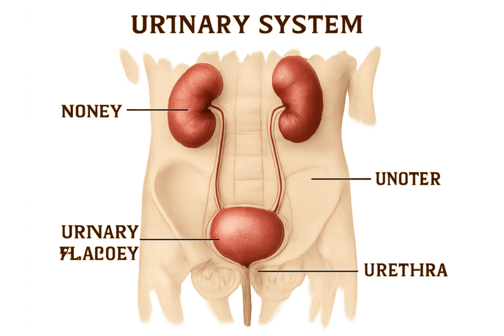
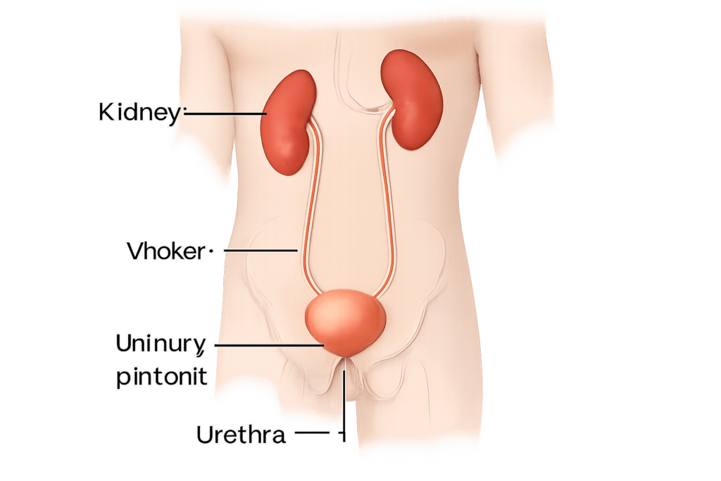
When you set out to correctly label the following components of the urinary system, focus on these primary organs:
- Kidneys
- Ureters
- Bladder
- Urethra
Each plays a unique role. Let’s break down what they do, and how to identify or label them on a diagram or in an exam setting.
1. Kidneys
The kidneys are two bean-shaped organs located on either side of your spine, roughly at the waist level. Their job is to filter waste and extra fluid from your blood, forming urine in the process. When labeling, place the kidneys near the upper back area — left and right.
Key Points:
- Often colored dark red/purple on diagrams
- Positioned towards the back of the abdominal cavity
- Filter blood, regulate fluid balance, and produce urine
2. Ureters
Ureters are thin tubes (one from each kidney) that transport urine from the kidneys to the bladder. Correctly label them as narrow tubes running downward from each kidney toward the pelvis.
Key Points:
- Each kidney has one ureter
- Shown as thin, pale lines or tubes connecting kidney to bladder
- Carry urine reliably via peristalsis (muscle contractions)
3. Bladder
The bladder is a hollow, muscular organ located in your lower abdomen. Its main purpose is to temporarily store urine until you’re ready to release it. On diagrams, label the bladder as a balloon-like structure in the lower pelvic area.
Key Points:
- Central, low in the pelvis
- Can expand and contract as it fills or empties
- Composed of muscle (detrusor muscle)
4. Urethra
Finally, the urethra is the tube that allows urine to exit the bladder and leave the body. When labeling, locate the urethra as the tube coming out of the bladder leading to the outside.
Key Points:
- Shorter in females, longer and curved in males
- Releases urine during urination
- Also conveys semen in males
Tips for Learning and Labeling the Urinary System
- Visualize with Diagrams: Use clear anatomical diagrams and practice labeling each part.
- Check Direction: Follow the urine’s path — from kidney to ureter, bladder, and finally out through the urethra.
- Mnemonic Devices: Phrases like “KUBU: Kidneys, Ureters, Bladder, Urethra” help reinforce sequence and structure.
Pros and Cons of Labeling Practice
Pros:
- Builds confidence for anatomy exams
- Strengthens understanding of body systems
- Useful for health and medical careers
Cons:
- Diagrams can vary in complexity
- Some anatomical differences exist (especially in urethra length by gender)
Final Thoughts
If you need to correctly label the following components of the urinary system, focus on clarity, sequence, and relative position: kidneys, ureters, bladder, and urethra. With regular practice, labeling these structures becomes second nature, supporting both academic and practical knowledge of human biology.